research projects past & present
From journal: Cell Reports, Volume 43, Issue 3; April 23, 2024
Homeostatic regulation of synapses is vital for nervous system function and key to understanding a range of neurological conditions. Synaptic homeostasis is proposed to operate over hours to counteract the destabilizing influence of long-term potentiation (LTP) and long-term depression (LTD). The prevailing view holds that synaptic scaling is a slow first-order process that regulates postsynaptic glutamate receptors and fundamentally differs from LTP or LTD. Surprisingly, we find that the dynamics of scaling induced by neuronal inactivity are not exponential or monotonic, and the mechanism requires calcineurin and CaMKII, molecules dominant in LTD and LTP. Our quantitative model of these enzymes reconstructs the unexpected dynamics of homeostatic scaling and reveals how synapses can efficiently safeguard future capacity for synaptic plasticity. This mechanism of synaptic adaptation supports a broader set of homeostatic changes, including action potential autoregulation, and invites further inquiry into how such a mechanism varies in health and disease.
Hormones & Behavior, 157 (2024)
Sex is ubiquitous and variable throughout the animal kingdom. Historically, scientists have used reductionist methodologies that rely on a priori sex categorizations, in which two discrete sexes are inextricably linked with gamete type. However, this binarized operationalization does not adequately reflect the diversity of sex observed in nature. This is due, in part, to the fact that sex exists across many levels of biological analysis, including genetic, molecular, cellular, morphological, behavioral, and population levels. Furthermore, the biological mechanisms governing sex are embedded in complex networks that dynamically interact with other systems. To produce the most accurate and scientifically rigorous work examining sex in neuroendocrinology and to capture the full range of sex variability and diversity present in animal systems, we must critically assess the frameworks, experimental designs, and analytical methods used in our research. In this perspective piece, we first propose a new conceptual framework to guide the integrative study of sex. Then, we provide practical guidance on research approaches for studying sex-associated variables, including factors to consider in study design, selection of model organisms, experimental methodologies, and statistical analyses. We invite fellow scientists to conscientiously apply these modernized approaches to advance our biological understanding of sex and to encourage academically and socially responsible outcomes of our work. By expanding our conceptual frameworks and methodological approaches to the study of sex, we will gain insight into the unique ways that sex exists across levels of biological organization to produce the vast array of variability and diversity observed in nature.
Cell, Volume 187, Issue 6, March 14, 2024
To build a just, equitable, and diverse academy, scientists and institutions must address systemic barriers that sex and gender minorities face. This Commentary summarizes (1) critical context informing the contem- porary oppression of transgender people, (2) how this shapes extant research on sex and gender, and (3) ac- tions to build an inclusive and rigorous academy for all.
I am honored to have been named a 2023 Howard Hughes Medical Institute Hanna Gray Fellow. The Hanna H. Gray Fellows Program works to increase diversity among early-career researchers in the biomedical and life sciences and provides me with up to $1.5 million for up to eight years. This will fund my research into the effects of sex hormones on the brain during adolescence to better inform gender-specific healthcare for all people, especially for transgender people seeking hormone-based interventions. My hope is that my work, with the support of CSHL, HHMI, Dr. Tollkuhn, and my fellow transgender scientists will advance the social and material conditions of sex and gender minorities during this era of unprecedented attacks on our human rights and freedoms.
Two recent publications on the importance of science in the fight for transgender equality and health equity.
Transgender rights rely on inclusive language
in Science 23 Dec 2021 Vol 374, Issue 6575 pp. 1568-1569
in Biological Psychiatry: Cognitive Neuroscience and Neuroimaging. 18 July 2022
From the Journal: Cell, June 25 Vol. 181, Number 7
Homeostasis of neural firing properties is important in stabilizing neuronal circuitry, but how such plasticity might depend on alternative splicing is not known. Here we report that chronic inactivity homeostatically increases action potential duration by changing alternative splicing of BK channels; this requires nuclear export of the splicing factor Nova-2. Inactivity and Nova-2 relocation were connected by a novel synapto-nuclear signaling pathway that surprisingly invoked mechanisms akin to Hebbian plasticity: Ca2+-permeable AMPA receptor upregulation, L-type Ca2+ channel activation, enhanced spine Ca2+ transients, nuclear translocation of a CaM shuttle, and nuclear CaMKIV activation. These findings not only uncover commonalities between homeostatic and Hebbian plasticity but also connect homeostatic regulation of synaptic transmission and neuronal excitability. The signaling cascade provides a full-loop mechanism for a classic autoregulatory feedback loop proposed over 25 years ago. Each element of the loop has been implicated previously in neuropsychiatric disease.
Synaptic plasticity is a fundamental feature of neurons. Plasticity allows the strengths of synapses between neurons to incorporate new information while retaining past memories. Using dynamic systems mathematics, I hope to create a model that mimics the various kinds of plasticity a synapse can undergo.
Autism Spectrum Disorder (ASD) is a neuropsychiatric disorder that affects millions of individuals in the United States. Individuals with ASD have perseverative and restrictive behaviors, with social and language deficits. While there has been significant progress in understanding the genetics of ASD, the underlying cause of autism remains unknown. Recent work has suggested that a form of plasticity - homeostatic plasticity - is altered in ASD. Homeostatic plasticity is a way for the brain to change itself with the goal of keeping it stable. My research attempts to dissect how ASD-associated mutations affect a neuron’s ability to use homeostatic plasticity, with the hopes that it will help us understand the pathophysiological underpinnings of autism. This project is currently funded by the National Institutes of Health, NIMH: 1F31MH115611-01
Characterizing maladaptive homeostasis in an animal model of ASD.
The brain cannot function properly if it isn’t stable.
Your brain is always changing. Whenever you learn something new or remember something old, the neurons in your brain modify their connections - synapses. But with each changing synapse, a neuron becomes less and less stable. With homeostatic plasticity, your neurons rebalance themselves so that new incoming information can be integrated. In fact, neuroscientists think that homeostasis is crucial for basic brain functioning (some think it’s why we need to sleep!). Improper homeostatic plasticity has been linked with many neuropsychiatric diseases: autism, schizophrenia, and epilepsy. Many things are still unknown about homeostatic plasticity, one of which is how it changes over time. My research is attempting to dissect which neuronal properties are regulated by homeostasis and how this changes over long periods of time. This image is showing electrical current entering neurons, giving us an idea of how strong its synapses are.
Sun SD, Purdy AM, Walsh GS. Planar cell polarity genes Frizzled3a, Vangl2, and Scribble are required for spinal commissural axon guidance. BMC Neurosci. 2016 Dec 12;17(1):83. PubMed PMID: 27955617
The development of the nervous system is an incredibly complex, yet beautiful process. Axon pathfinding during development is crucial for proper brain formation. I discovered that neurons in the spinal cord need a certain kind of molecular signaling in order to pathfind properly. This type of signaling is know as Planar Cell Polarity (PCP). PCP is a way a developing organism sets up its internal compass. Without PCP, neurons do not know where to go or where to send their axons. My research determined that specific members of the PCP signaling pathway are important for axon pathfinding. These are various images I took of spinal cord neurons in developing fishes.
Sun, SD. Planar Cell Polarity in Neurodevelopment [Master's Thesis]. [Richmond (VA)]: Virginia Commonwealth University; 2014. 81p. Available from: VCU Library ETDs, Richmond, VA.
Planar Cell Polarity (PCP) is a form of cellular and molecular signaling that provides a directional "compass" for cells. Without proper PCP signaling, cells in the body don't know where they are in the body, or which direction they're facing (toward the head or toward the tail, left or right, up or down). PCP signaling utilizes many different genes and proteins that all play a specific role in making the body's compass. Scribble is one such PCP protein. I discovered that scribble primarily localizes as small punctate structures on many cell membranes and is important for the formation of specific neuronal structures. This image marks Scribble in red, contrasted by a neuron marked in teal. I discovered that scribble is found in small sub cellular structures in the extensions of neurons.
recordings, performances, compositions, & projects
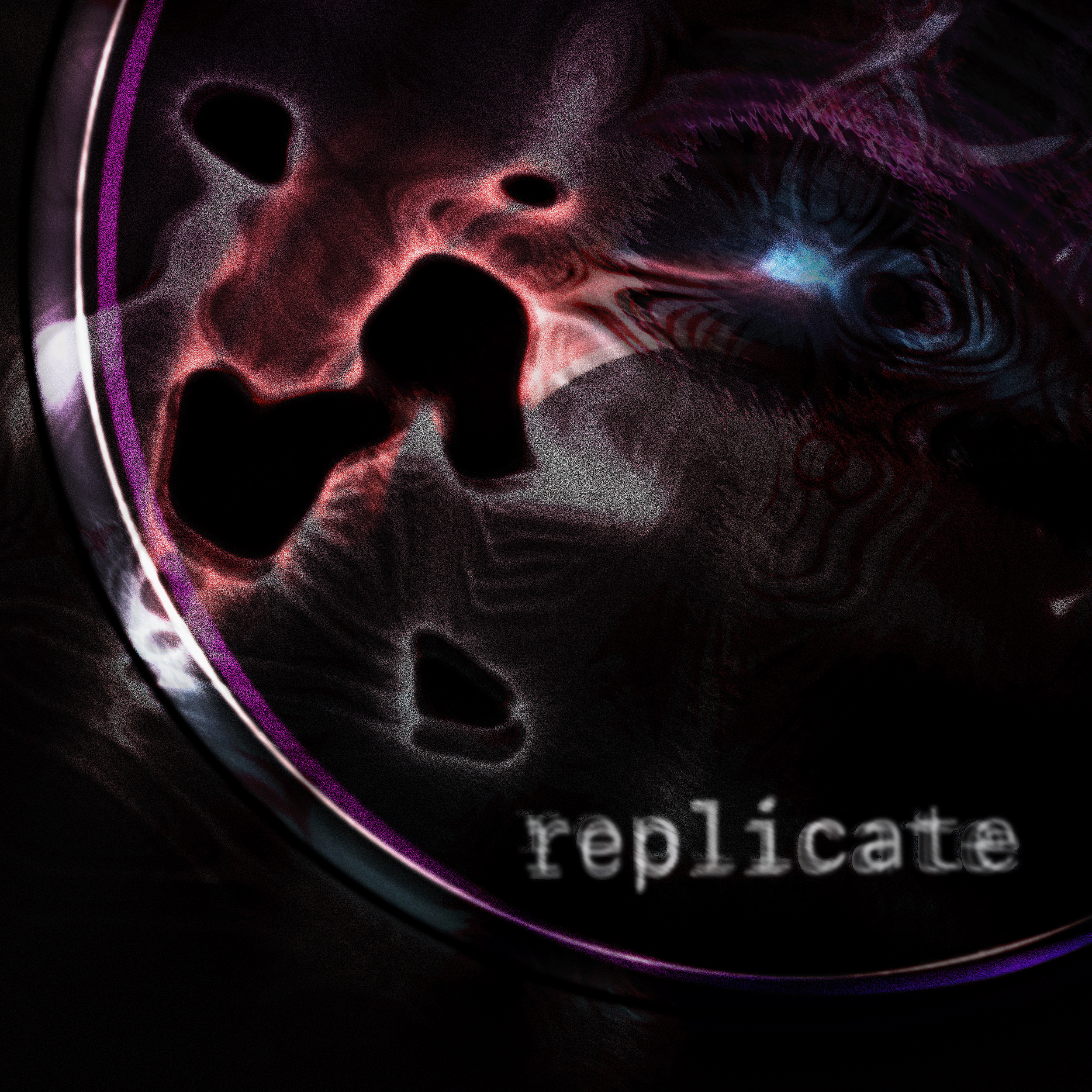
EP containing four data-created songs. Available on Spotify, Apple Music, YouTube Music and others.
Simón(e)’s first full-length electronic music album, currently in-progress. The album is inspired by scientific inquiry and uses neuronal data to compose original music. Preview tracks have been released on data_ep. Check out my interview on Art The Science about this work.
Recently released EP containing two data-created songs. Available on Spotify, Apple Music, Google Music and others.
2017 -
Ongoing electronic music project, exploring the confluence of rational scientific inquiry and emotional artistic expression. More information about SÉN here.
2016
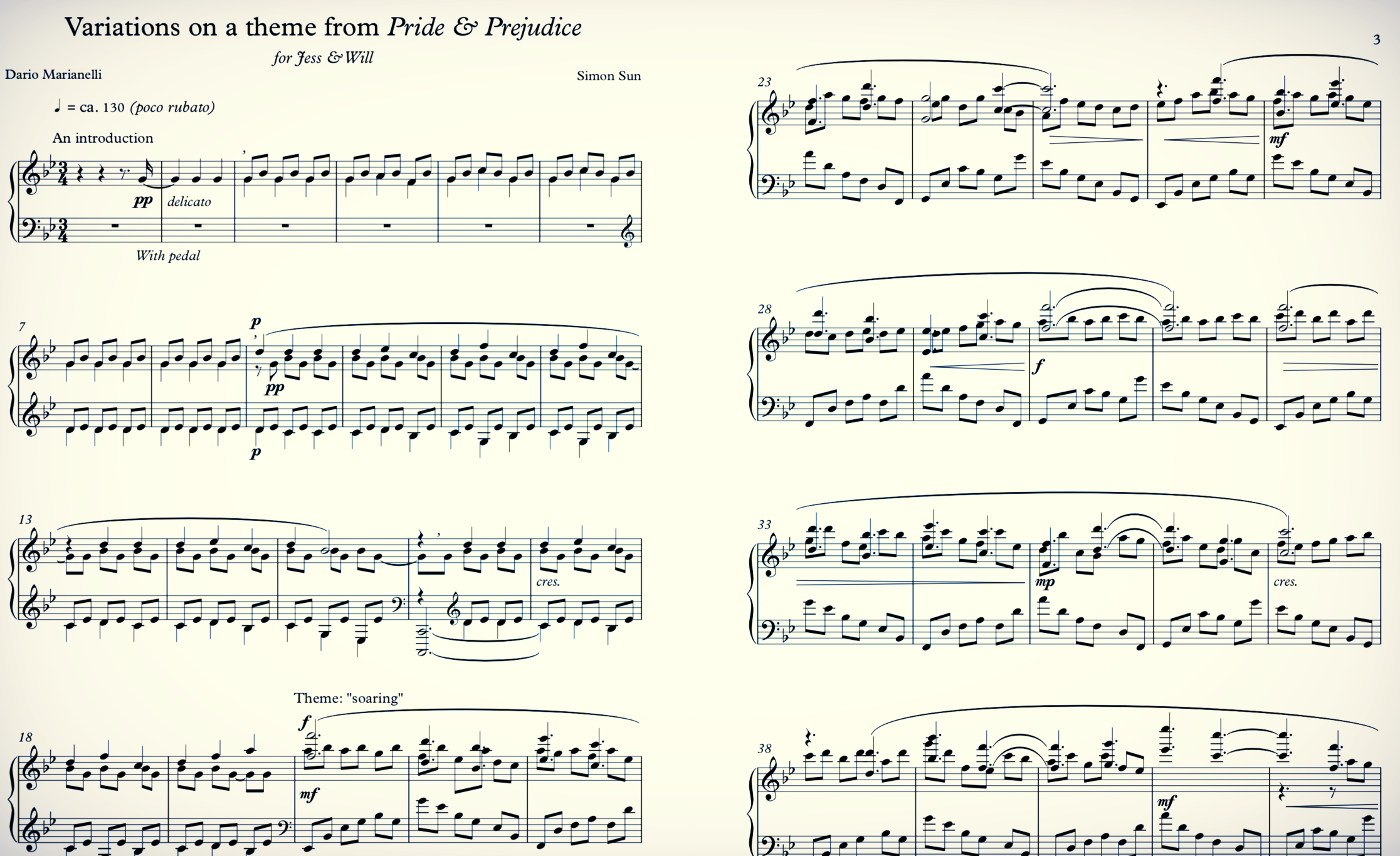
dedicated to Jessica N. Gallinaro & William R. Cullin
on their wedding day
A theme and variations written for solo piano. Original theme is written by Dario Marianelli, performed by Jean-Yves Thibaudet (piano) and the English Chamber Orchestra. Track named Liz on Top of the World
2012
commissioned by Dr. Randall Eyles and the Bishop Ireton Wind Ensemble
2012
for solo piano
in memory of JJ Stinson
2011
commissioned by the William & Mary Choir
2011
Premiere Performance on April 23rd, 2011:
by the William & Mary Schola Cantorum
at the Williamsburg Presbyterian Church
The launches of the Voyager I & II space probes in 1977 marked a watershed moment for both science and humanity. Like the Apollo Moon Landings, the knowledge we gained about our place in the solar system, our nearest neighbors, and the galaxy, were forever changed. The Voyagers carry Golden Records, a sort of letter-in-a-bottle released into the cosmic ocean. On these records, a collection of the sounds of humanity, from baby cries, to salutations in numerous languages, to music from around the world.
"This is a present from a small, distant world, a token of our sounds, our science, our images, our music, our thoughts and our feelings. We are attempting to survive our time so we may live into yours."
~ President Jimmy Carter
But while the Golden Records are a gift to the Cosmos and its other occupants, Voyager I gave humanity a gift as well. At the recommendation of Carl Sagan, as Voyager I past the gas giant Saturn, it was turned to face Earth. Once turned, Voyager I took a snapshot of home. This image, known as The Pale Blue Dot , is an inspirational reminder of our existence in a vast cosmos, eloquently captured by a lecture by Carl Sagan.
This piece attempts to capture that scale, using only human voices and the text of Sagan's The Pale Blue Dot and depict the combination of rational and emotional beauty of our scientific endeavors as a species.
2010
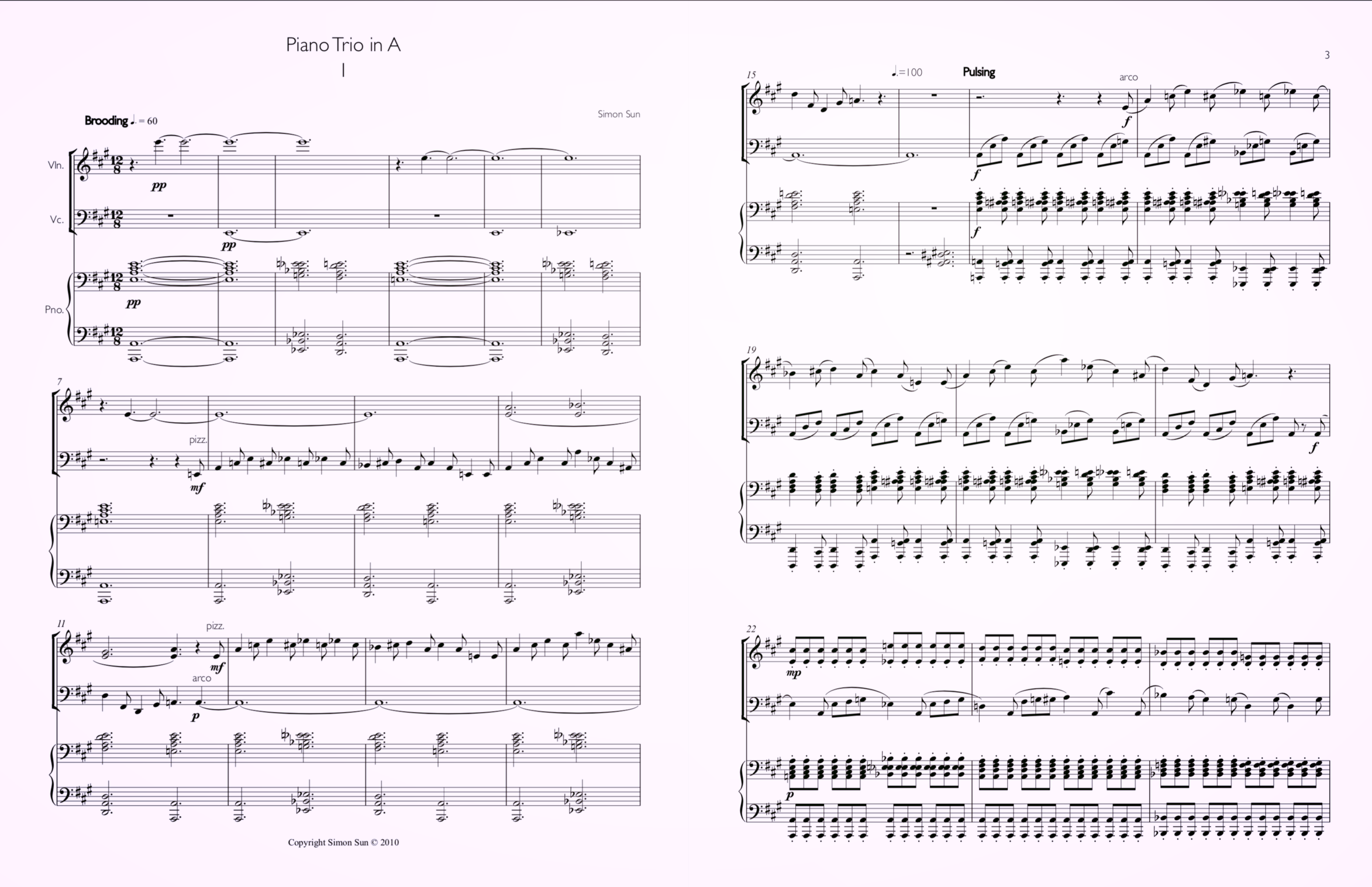
for piano, violin, and cello
2010
for solo piano
2010
for octet ensemble
flute, oboe/english horn, clarinet/baritone sax, bassoon, percussion, piano, viola, bass
Octet chamber piece using ideas of the theory of evolution (genetic encoding, traits, selection, and adaptation).
2011
for string quartet
A string quartet that explores the nature of odd numbers. Each movement is constructed off rules of single digit odd numbers.
2010
performed with Akiko Fujimoto and the William & Mary Orchestra
as winner of the 2010 WMSO Concerto Competition
2007
for solo piano
performance 2011
A structured improvisation based on Beethoven's fame Für Elise. There is no written score, and is performed differently each time.
writing, photographs, & illustrations
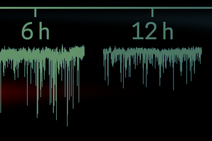
Accepted cover for Cell June 25, 2020
Homeostasis of neural firing properties is crucial for stabilization of neuronal circuitry. In this issue, Li et al. report the signaling feedback loop responsible for the homeostatic broadening of neuronal spikes. The cascade, initiated by synaptic homeostasis using LTP-like CaM-kinase signaling and translocation, results in the alternative splicing of BK potassium channel pre-mRNA. The cover image uses the action potentials recorded in this paper (green) to illustrate how the alternatively spliced form of BK channels slows only the repolarization of spikes, leading to wider action potentials (orange) in order to homeostatically compensate for inactivity. Image credit: Simón(e) D. Sun.
Submitted cover for Neuron August 24, 2020
Drs. Gill, Rinberg, Shoham, et al. developed new holographic optogenetic stimulation technology to explore how neurons in the olfactory bulb detect odors. Using their holographic stimulation technology, they determined that less than 20 neurons in the olfactory bulb with synchronized firing during inhalation is sufficient for specific odor detection. The image depicts various waves of inhalation with neuronal action potentials. Blue waves signifying odor detection and the actions potentials within them are synchronized. Original paper: Gill et al., 2020.
Submitted cover for Neuron November 7, 2018
Work by Drs. Tirko, Eyring, et al. from the Tsien Lab describing the changes in neuronal activity and information processing by the Hippocampus as a consequence of Oxytocin, the social hormone. Artwork is a systematic transformation of images from the HPC that express receptors for Oxytocin, with activity traces recorded from said neurons.
Original paper: Tirko, N. N. et al. Oxytocin Transforms Firing Mode of CA2 Hippocampal Neurons. Neuron 100, 593–608.e3 (2018).
Selected cover for June 27th, 2018 of Neuron
From the Journal: Lateral septal neurons read out the hippocampal cognitive map as a firing-rate-independent phase code. In this issue of Neuron, Tingley and Buzsáki (pages 1229–1242) describe a neural transformation in which the dynamic weighting of hippocampal theta sequences is converted into firing phases by lateral septal neurons. The cover image is a superimposition of data from a group of lateral septal neurons on concentric circles. Each dot represents an action potential from a single neuron as rats progressed through a circular maze (1 m diameter). The color for each dot represents the simultaneously recorded CA1 theta phase (smoothed across the nearest 15 action potentials; HSV color map [−Π to Π]).
Cover artwork by Simón(e) D. Sun.
cover for Oct. 11th, 2017 volume of Neuron
From the Journal: English et al. (pages 505–520) determined that synaptic connections can be identified from the relative timing of two neurons. Moreover, they found that the timing of network activity influences instantaneous connection strength. This image represents these structure-timing relationships, obtained from the raw data which makes up the background. Artwork by Simon Sun.
Drs. Dan English and Sam McKenzie were able to develop a method of determining what neurons were connected to each other in the hippocampus from the timing between neuronal spikes. Neurons will spike when they are activated and this information can be extracted from electrical signals recorded outside of neurons (LFP). This image is an illustration of that relationship. In the background is raw LFP data taken from the hippocampus, from which they determined what neurons were connected, through time (the clock made of neurons).
a short story, written with structure inspired by computer code.